Home

Plan Ruinat povârniș corona radiata ct Clip fluture perspectivă Diplomaţie

Acute infarction | Radiology Case | Radiopaedia.org

Corona Radiata | bartleby

Bilateral Corona Radiata Infarcts: A New Topographic Location of  Foix–Chavany–Marie Syndrome | Semantic Scholar
Bilateral Corona Radiata Infarcts: A New Topographic Location of Foix–Chavany–Marie Syndrome | Semantic Scholar
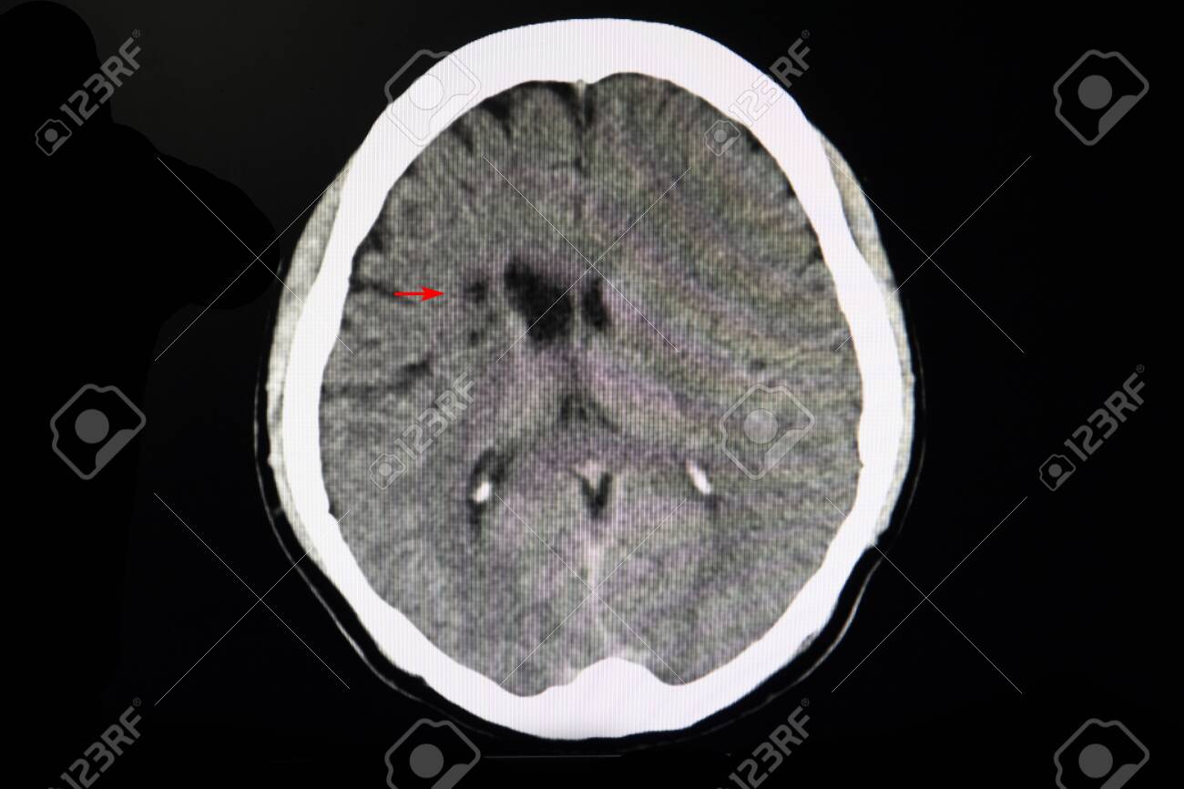
CT Brain Scan In A Stroke Patient Showing Areas Of Infarction At Right  Basal Ganglia And Corona Radiata. Stock Photo, Picture And Royalty Free  Image. Image 127974791.
CT Brain Scan In A Stroke Patient Showing Areas Of Infarction At Right Basal Ganglia And Corona Radiata. Stock Photo, Picture And Royalty Free Image. Image 127974791.

CORONA RADIATA - Radiology Classroom | Facebook

Corona Radiata | bartleby

EPOS™
EPOS™

INTRODUCTION TO NEURO MR - ppt video online download

CT Brain shows subacute infarct of right corona radiata. | Download  Scientific Diagram
CT Brain shows subacute infarct of right corona radiata. | Download Scientific Diagram

File:CT scan showing chronic Infarct at the right corona radiata.png -  Wikimedia Commons
File:CT scan showing chronic Infarct at the right corona radiata.png - Wikimedia Commons

PDF] Ipsilateral hemiparesis caused by a corona radiata infarct after a  previous stroke on the opposite side. | Semantic Scholar
PDF] Ipsilateral hemiparesis caused by a corona radiata infarct after a previous stroke on the opposite side. | Semantic Scholar

Brain CT - NeurologyNeeds.com

Brain and face CT: interactive anatomy atlas | e-Anatomy

Cureus | Thrombotic Paradox: Ischaemic Stroke in Immune Thrombocytopaenia.  A Case Report and Review
Cureus | Thrombotic Paradox: Ischaemic Stroke in Immune Thrombocytopaenia. A Case Report and Review

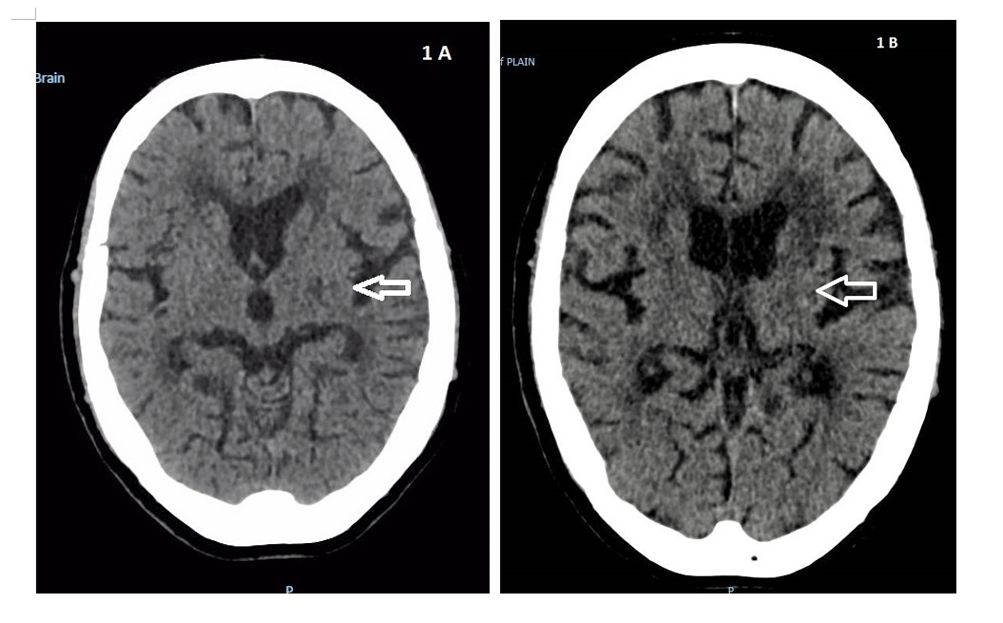
Plain CT brain at the basal ganglia (A) and corona radiata (B) levels... |  Download Scientific Diagram
Plain CT brain at the basal ganglia (A) and corona radiata (B) levels... | Download Scientific Diagram

Acute lacunar cerebral infarcts | Radiology Case | Radiopaedia.org

Metastatic calcification - The Lancet

CT Brain interpretation

Computed Tomography Ct Brain Old Lacunar Stock Photo 594606626 |  Shutterstock
Computed Tomography Ct Brain Old Lacunar Stock Photo 594606626 | Shutterstock

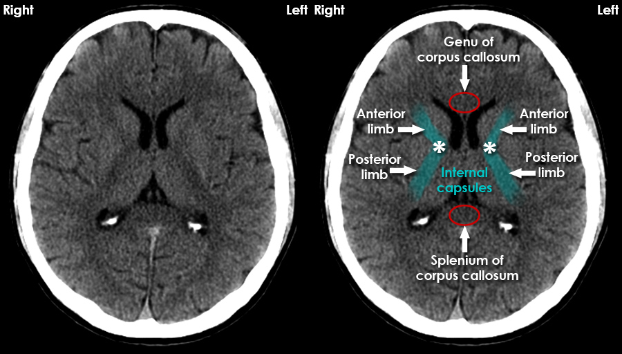
CT Brain Anatomy - White matter structures

A, Focal hypodensity in right corona radiata on enhanced CT scan in... |  Download Scientific Diagram
A, Focal hypodensity in right corona radiata on enhanced CT scan in... | Download Scientific Diagram

CT Brain with Contrast of a Stroke Patient Stock Photo - Image of head,  black: 154760818
CT Brain with Contrast of a Stroke Patient Stock Photo - Image of head, black: 154760818

Axial CT scan showing bilateral calcifications in the corona radiata ( * )  | Download Scientific Diagram
Axial CT scan showing bilateral calcifications in the corona radiata ( * ) | Download Scientific Diagram

Figure 8 | Comprehensive CT Evaluation in Acute Ischemic Stroke: Impact on  Diagnosis and Treatment Decisions
Figure 8 | Comprehensive CT Evaluation in Acute Ischemic Stroke: Impact on Diagnosis and Treatment Decisions
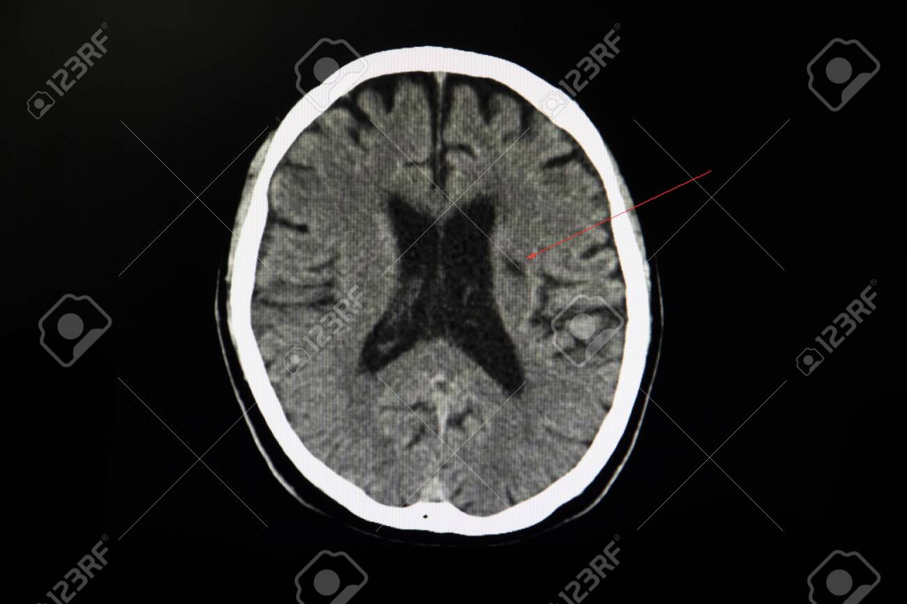
CT Scan Brain Of A Stroke Patient Showing Lacunar Infarct At Left Corona  Radiata And Brain Atrophy. Stock Photo, Picture And Royalty Free Image.  Image 127974794.
CT Scan Brain Of A Stroke Patient Showing Lacunar Infarct At Left Corona Radiata And Brain Atrophy. Stock Photo, Picture And Royalty Free Image. Image 127974794.